An Unusual Saltwater Bead Cultured Pearl
Figure 1. Left: This gourd-shaped saltwater bead cultured pearl (2.04 g and 15.90 mm high) displays two different colors. Right: Magnification indicates that the two parts grew together naturally. Photos by Yanhan Wu.
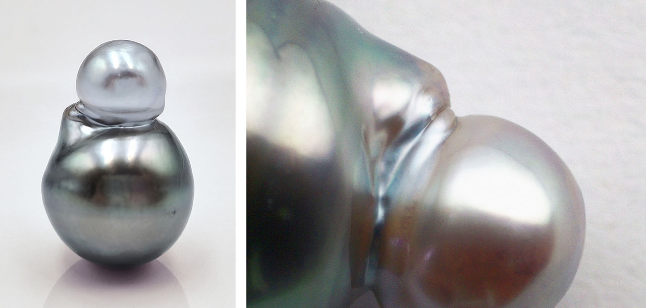
A saltwater pearl with an unusual appearance was discovered in the pearl market in the city of Zhuji, in China’s Zhejiang Province. It was shaped like a gourd, consisting of a silver gray part and a black part with green overtone (figure 1, left). The pearl’s unusual color and shape raised suspicions about whether it had been artificially processed.
Figure 2. Left: The UV-Vis-NIR reflection spectra of the two parts of the pearl showed the same absorption positions and differences in intensity. Spectra are offset vertically for clarity. Right: Raman spectroscopy displayed the bands of aragonite and organic components.

No silver was detected with energy-dispersive X-ray fluorescence in either part, indicating that the pearl had not been treated with silver nitrate dye. The ultraviolet/visible/near-infrared (UV-Vis-NIR) reflectance spectra of both parts showed absorptions at 279, 405, 500, and 700 nm (figure 2, left), consistent with previous studies on the absorption of saltwater cultured pearls (S. Karampelas et al., “UV-Vis-NIR reflectance spectroscopy of natural-color saltwater cultured pearls from Pinctada Margaritifera,” Spring 2011 G&G, pp. 31–35). In the visible light region, only the cause of the 405 nm band has been identified; it is attributed to a kind of porphyrin called uroporphyrin (Y. Iwahashi et al., “Porphyrin pigment in black-lip pearls and its application to pearl identification,” Fisheries Science, Vol. 60, No. 1, 1994, pp. 69–71). The black part was more absorbed than the gray part due to its richer color.
Figure 3. 3D fluorescence spectra of the black part of the pearl (left) and the gray part (right).
Under long-wave ultraviolet radiation, both parts exhibited purplish blue fluorescence. However, the gray part exhibited significantly more intense fluorescence than the black part. The 3D fluorescence spectra confirmed this. The strongest fluorescence in the black part was at 336 nm (figure 3, left), while the strongest in the gray part was at 340 nm (figure 3, right). The fluorescence intensity between the two differed by more than 2.5 times. The gray part still had weak fluorescence centers at 432 nm, while the black part did not.
Figure 4. μ-CT scan images of the saltwater bead cultured pearl at different depths.
The X-ray computed microtomography (μ-CT) scan in figure 4 shows that the outer layer of the two parts was connected by nacre, while the inner layer was composed of voids and organic components. Both parts had a distinct bead nucleus. The organic matter between the bead nucleus and the nacre had a dark gray circular shape. The nacre thickness in both parts was 3–4 mm.
Many mechanisms combine to affect the color of pearls during their growth process (Z. Wang et al., “How cultured pearls acquire their colour,” Aquaculture Research, Vol. 51, 2020, pp. 3925–3934), making it difficult to pinpoint the exact mechanism. This research can be used to further study the relationship between organic compounds and the color and fluorescence reactions of saltwater pearls, excluding variables such as mollusk type and environmental factors.
Leave a Reply
Want to join the discussion?Feel free to contribute!